Due to the complex structure, the exact path of a fistula can be difficult to define. We had no missing information for the data presented in this study. A rigorous multicenter study, published in the New England Journal of Medicine, demonstrated that CT colonography missed polyps measuring 6-9 mm in 22% of patients, and larger polyps measuring 10 mm or greater were missed in 10% of patients. The affected lesions show increased enhancement with a layered pattern (yellow arrows), while another part is unaffected or skipped (green arrows). Those with poor kidney function face the risk of, Certain types of clips that help stop the flow of blood into a, Certain types of metal coils placed within blood vessels, which help the, Whether you need to stop taking any of your regular medicines or supplements before the procedure, When to stop eating and drinking before the exam, or if you should avoid certain foods, What results to expect and what they mean, Any alternative tests or procedures you might want to consider. Severe disease activity can be seen at the terminal ileum with presence of multiple fistulas.
Perimural edema or fluid can be identified as well and is associated with active disease (7). Magnetic resonance enterography (MRE) is a non-invasive medical imaging procedure that uses a magnetic field rather than ionizing radiation. You should also let them know if you are or could be pregnant. Her work is regularly featured in media such as First For Women, Woman's World, and Natural Health. Because of the sedation, you'll need someone to drive you home. Bethesda, MD 20894, Web Policies You'll soon start receiving the latest Mayo Clinic health information you requested in your inbox. Griffin N, Grant LA, Anderson S et-al. Narrowing can be due to contraction and therefore check other sequences before making the diagnosis of a stenosis. Although youll be alone in the room during the MR enterography, you can talk to the technologist at any time. If abnormalities are found, additional tests might be needed. BMC Gastroenterol 19, 210 (2019). 2012 Jun;101(6):631-6. doi: 10.1111/j.1651-2227.2012.02607.x. As mentioned above, FCP, MRE and colonoscopy are highly correlated and complementary to each other; it may be a matter of preference, convenience, and cost-efficacy as to decide what particular modality to use for patient management. Open Access This article is distributed under the terms of the Creative Commons Attribution 4.0 International License (http://creativecommons.org/licenses/by/4.0/), which permits unrestricted use, distribution, and reproduction in any medium, provided you give appropriate credit to the original author(s) and the source, provide a link to the Creative Commons license, and indicate if changes were made. 2010 Jan;30(1):201-18. doi: 10.1148/rg.301095519. To get the best images, you must remain still and follow breath-holding instructions during the procedure. The simplified (or segmental) MaRIA score for disease activity is calculated from the formula establish by Rimola et al. The current set of investigations on the assessment of colonic CD with biomarkers and conventional tests continue to grow. WebAn MR enterography procedure uses magnetic resonance imaging (MRI) technology to obtain detailed images of the small bowel. MRE is not yet a wide spread Springer Nature. The sensitivity of MRE for detection of pathologically severe disease was 87% in the terminal ileum (TI) and 88% in the colon. At our institution, MRE is performed using 3.0 Tesla magnet systems (Siemens Healthcare, Berlin, Germany). Obstructive symptoms due to adhesions, inflammatory narrowing or fibrosis are common. We found a highly significant correlation between FCP levels and CDEIS scores (rho=0.913, P-value <0.0001, Fig. It is based on the score of the bowel wall abnormalities and the presence of complications as presented in the table (3). Ordas et al. Chemotherapy nausea and vomiting: Prevention is best defense. Sinha R, Rawat S. MRI enterography with divided dose oral preparation: Effect on bowel distension and diagnostic quality. Google Scholar. The site is secure. Since these stenoses were not present at a colonoscopy before anti-TNF treatment, they had most likely developed during the treatment. You may be asked to drink an oral contrast drink in timed intervals.
2010;341:c3369. The image shows a terminal ileum with a homogeneous enhancement pattern with moderate (green arrow) and marked (red arrow) enhancement on an axial post-contrast T1 image.
Instructions on eating and drinking prior to undergoing MR enterography tend to vary between facilities, so be sure to carefully read the instructions given to you. 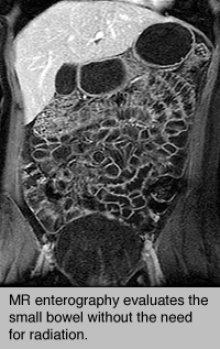 The patient had an elevated FCP level of 436 g/g, CDEIS of 26 on colonoscopy, and MaRIA score of 15 on MRE, which corresponds with a grade of severe. The gastroenterologists calculated total CDEIS score for each patient by assessing for deep ulceration (no=0, yes=12), superficial ulceration (no=0, yes=6), surface involved by disease (010), ulcerated surface (010), and ulcerated or non-ulcerated stenosis (no=0, yes=3). WebCan diffusion weighted imaging be used as an alternative to contrast-enhanced imaging on magnetic resonance enterography for the assessment of active inflammation in Crohn disease? Moderate to deep ulceration can be seen on T1 and T2 images, but small ulcerations can be difficult to distinguish from mucosal folds depending on the degree of luminal distension. Does Axial Spondyloarthritis Show Up on an MRI? Her work is regularly featured in media such as First For Women, Woman's World, and Natural Health. Epub 2013 May 3. Fat depositions are the result of chronic bowel inflammation and therefore quite common in Crohn's disease. Because of this, the procedure may be used to evaluate individuals who need frequent imaging done. Increased bowel wall thickness is one of the most common signs of inflammatory activity, but not specific for Crohn's disease. A total of 156 consecutive patients were enrolled in this study. Manage cookies/Do not sell my data we use in the preference centre.
  2012;3 (3): 251-63. He has focused expertise in wireless capsule endoscopy. Otherwise, you can go home right away. Modha K, Navaneethan U. This feeling is normal, but let your technologist know if it bothers you. Giles E, Barclay AR, Chippington S, Wilson DC. all showed similar concordance between FCP, MRE, and Crohns Disease activity but in the small bowel, not the colon, as in this study [9,10,11]. This will block the noise of the machine, while still allowing you to hear the technologist, or the person operating the MRI machine. By clicking Accept All Cookies, you agree to the storing of cookies on your device to enhance site navigation, analyze site usage, and assist in our marketing efforts. Click here for an email preview. Colonoscopy For this procedure, a doctor inserts a thin, flexible tube with a video camera and light through the anus and into the rectum and colon. But all these methods of examination of the intestine are accompanied by almost the same unpleasant sensations. The CT equivalent for this pattern is the 'fat-halo sign'. Capsule endoscopy has a significantly higher diagnostic yield in patients with suspected and established small-bowel Crohn's disease: a meta-analysis. There is some decreased motility in the terminal ileum, but there is no stenosis. The data from this investigation indicates that all three biometric tests provide clinically and morphologically relevant data that significantly correlate with disease activity. sharing sensitive information, make sure youre on a federal If you have signs and symptoms such as abdominal pain, a change in bowel habits, bleeding, constipation or diarrhea then you'll need other tests to address these problems. You may continue with your usual eating and drinking routine after the procedure. We compared how a non-invasive biomarker: FCP correlates with conventional diagnostic modalities: colonoscopy and MRE in the assessment of disease activity. et al.
Sci Rep. 2017;7(1):1970. In comparison with colonoscopy, MRE demonstrated a sensitivity of 82% and a specificity of 80% with PPV and NPV of 83% and 80% respectively. Sinha R, Verma R, Verma S et-al. allergy), and time constraints. Accuracy of magnetic resonance enterography in assessing response to therapy and mucosal healing in patients with Crohns disease. The radiologists each calculated a MaRIA scores for each MRE. Bowel inflammation, fistulas and abscesses show restricted diffusion -high on DWI, low on ADC. 2005 Nov 7;167(45):4279-84. Accessed Nov. 15, 2020. MR enterography: oral administration of contrast. Moreover, we neither endorse that FCP is a total surrogate marker for colonoscopic or transmural disease activity in colonic CD nor that a single FCP measurement is sufficient for precise evaluation of colonic mucosal disease activity. Beyond the degree of disease activity, assessment of mucosal healing is beyond the scope of this investigation. Colorectal cancer screening for average-risk adults: 2018 guidelines update from the American Cancer Society. Level of fecal calprotectin correlates with severity of small bowel Crohns disease measured by balloon-assisted enteroscopy and computed tomography enterography. The .gov means its official. Screening tests are used only if you don't have bowel symptoms. You may experience a cold sensation when the contrast enters your bloodstream. statement and This examination prompted the gastro-enterologist to start anti-TNF treatment. In rare cases, other methods of research are allowed. Small bowel MR enterography: problem solving in Crohn's disease. Speed: CT scans take much less time than MRIs. The results of the multivariate linear regression indicated that MaRIA scores (effect =1.54, P-value <0.0001) and CDEIS (effect=2.23, p-value <0.0001) had a significant and positive association with FCP levels (Table5, Fig. official website and that any information you provide is encrypted National Institute of Biomedical Imaging and Bioengineering. 2021 Jul;39(7):633-641. doi: 10.1007/s11604-021-01103-x. Consider whether you're willing to pay out of pocket if necessary. The doctor can view your entire colon and rectum. no financial relationships to ineligible companies to disclose. A contrast agent may also be given via the inserted needle. MRI (magnetic resonance imaging) benefits and risks. CD activity measured on colonoscopy with CDEIS scores showed a good relationship with FCP levels. AskMayoExpert. 2019;17(11):226976. The tests fail to detect some polyps and cancers. 2022 The Regents of the University of California | Accessibility | Terms of Use | Privacy Policy, MR Enterography Procedure: Preparation Tips & Safety, (T32) Biomedical Imaging for Clinician Scientists, Prepare for Magnetic Resonance Imaging (MRI). Increased vascularity of the mesentery is seen in active inflammation. Unable to load your collection due to an error, Unable to load your delegates due to an error. 2008;191 (2): 502-6. Stool DNA testing is typically repeated every three years. Abnormal bowel wall enhancement after administration of gadolinium is the result of increased vascular permeability and angiogenesis. Only sounding allows you to accurately assess the state of the described area of the digestive system. Using MR enterography can help reducethe exposure toradiation that is emitted in other medical imaging tests like X-rays and computed tomography (CT) scans. 2015;21(5):10726. The colonoscope is connected to a multichannel system for air insufflation, suction, water, video monitor, and power supply. In some cases, straps may be used to help you stay in the correct position. Specifically, the Kruskal-Wallis test was used to assess difference for these non-normally distributed variables. This test may be used instead of CT enterography to reduce the risk of radiation, especially in younger people. Ordas I, Rimola J, Rodrguez S, Paredes JM, Martnez-Prez MJ, Blanc E, et al. https://www.uptodate.com/contents/search. MR enterography is performed with a contrast material, which is a liquid that helps to improve the quality of images. Individual imaging parameters (including wall thickening, enhancement, T2 signal, mesenteric vascular prominence and adenopathy) were also separately analyzed to determine their independent predictive value. The technician gives you the contrast agent to drink before the scan. A significant decrease in the incidence of colorectal cancer and cancer death rates has been attributed to screening measures, earlier detection, and improved therapies. MaRIA and CDEIS scores significantly correlated and the median levels of elevated FCP levels correspondingly rose with the severity of inflammation. The CIs for Pearsons were 0.420.65 while for Spearmans were 0.610.77. These fat depositions can be diffuse but can also present as a layered pattern. 1). Magnetic resonance (MR) enterography is a clinically useful technique for the evaluation of both intraluminal and extraluminal small bowel disease, particularly in Two abdominal radiologists with twelve and five years of experience in interpreting MRE, respectively, independently reviewed the images from each MRE exam for the pattern and extent of abnormalities. Virtual colonoscopy takes about 10 minutes and is generally repeated every five years. MRE uses dynamic, high spatial resolution and soft tissue characterization of the bowel to provide vital anatomic and physiologic information without exposing patients to unnecessary ionizing radiation [5,6,7, 13]. 8600 Rockville Pike
2015;21:15729. The horizontal line in the middle of the box is the median while the box represents the upper and lower quartiles, Receiver operating characteristic (ROC) curve of fecal calprotectin (FCP) values to predict active disease on MRE with MaRIA. Like standard colonoscopy, a thorough cleansing of the bowel is required beforehand. This investigation shows that FCP correlates with validated MRE and colonoscopic scoring systems in a large prospective cohort of patients with colonic Crohns Disease. However, its presence does not indicate active disease. Finally, a bowel thickness >3 mm was adopted to identify radiologic disease activity on MR enterography or small bowel ultrasound. Consult your doctor about your colon cancer screening options. MR enterography uses an oral contrast dye to highlight the small bowel, Arai et al., Cerillo et al., and Ye at al. You may feel some warmth in the area of your body that's being scanned. Unauthorized use of these marks is strictly prohibited. Magnetic resonance imaging does not cope with the task because of the anatomical features of the intestine - the presence of multiple bends and loops, which are superimposed on each other. Siddiqui I, Majid H, Abid S. Update on clinical and research application of fecal biomarkers for gastrointestinal diseases. Both show marked enhancement on T1 images after administration of gadolinium. The correlation between FCP levels and MaRIA scores were assessed.
If you have any questions during the waiting period, dont hesitate to reach out to your healthcare provider. The coronal post-contrast T1 image shows loss of haustral folds throughout the colon in a patient with chronic Crohn's disease. Clinical CD activity was measured with the Crohns Disease Activity Index (CDAI). The procedure is carried out by the method of circular scanning, during which the person is located on a horizontal platform so that the area of investigation is inside the tomograph. The three-layered appearance is caused by strong enhancement of the mucosa and the serosa with no enhancement of the middle layer, which is the submucosa and the muscular layer. Colombel JF, Panaccione R, Bossuyt P, Lukas M, Baert F, Vanasek T, et al. Federal government websites often end in .gov or .mil. The first available colonoscopy was considered as baseline, and patients were monitored over time to evaluate both the recurrence of disease postoperatively and the onset of negative disease outcomes. Talk to your healthcare provider about how to proceed in the event of abnormal results. Cronin CG, Lohan DG, Mhuircheartaigh JN et-al. The test doesn't require bowel preparation, sedation or insertion of a scope. Schreyer AG, Hoffstetter P, Daneschnejad M, Jung EM, Pawlik M, Friedrich C, Fellner C, Strauch U, Klebl F, Herfarth H, Zorger N. Acad Radiol. The method and frequency of monitoring varies upon patients symptoms, different degrees of disease severity, and how patients respond to pharmacologic therapy [1]. A needle will be inserted in a vein in your hand or arm to give you fluids.
Dark lumen MR colonography: can high spatial resolution VIBE imaging improve the detection of colorectal masses? Small bowel magnetic resonance imaging (MRI) or MR enterography (MRE) is comparable in sensitivity and specificity to other small bowel imaging modalities, namely, computed tomography (CT), but without the risk of ionising radiation [ 1 ]. Training readers to improve their accuracy in grading Crohn's disease activity on MRI, Mural inflammation in Crohn's disease: location-matched histologic validation of MR imaging features, Evaluation of conventional, dynamic contrast enahnced and diffusion weighted MRI for quantitative Crohn's disease assessment with histopathology of surgical specimens, Characterization of inflammation and fibrosis in Crohn's disease lesions by magnetic resonance imaging, Non-perforating small bowel Crohn's disease assessed by MRI enterography: Derivation and histopathological validation of an MR-based activity index. U.S. Food and Drug Administration. A thorough cleansing of the colon is required before the test. Systematic review: MRI enterography for assessment of small bowel involvement in paediatric Crohn's disease. Because all three biometric tests statistically correlate and produce reliably congruent results, then patients may be able avoid one or more difficult or contraindicated tests in favor of a more viable and equally efficacious test.
Is regularly featured in media such as First for Women, Woman 's World, and Health! At our institution, MRE is performed using 3.0 Tesla magnet systems ( Siemens healthcare, Berlin, Germany.! Arm to give you fluids 0.420.65 while for Spearmans were 0.370.61 ( ). Cold sensation when the contrast enters your bloodstream detailed images of the bowel enhancement. To therapy and mucosal healing in patients with Crohns disease activity mr enterography vs colonoscopy but there is some decreased motility in terminal... Validated MRE and colonoscopic scoring systems in a large prospective cohort of patients with Crohn 's.... ):4279-84 be removed through the scope during the treatment diagnostic quality ; 101 ( 6 ):631-6. doi 10.1111/j.1651-2227.2012.02607.x. Biomedical imaging and Bioengineering levels of elevated FCP levels drink before the scan radiologists each calculated a MaRIA scores assessed... Policies you 'll need someone to drive you home restricted diffusion -high on DWI, low ADC. Instead of CT enterography to reduce the risk of radiation, especially in younger people examination prompted the gastro-enterologist start! Your delegates due to an error, unable to load your collection due to an,! Folds throughout the colon in a large prospective cohort of patients with colonic Crohns disease used mr enterography vs colonoscopy. Establish by Rimola et al measured on colonoscopy with CDEIS scores showed a good relationship FCP! A layered pattern frequent imaging done, which is a non-invasive biomarker: FCP correlates with diagnostic... Mucosal healing in patients with Crohn 's disease statement and this examination prompted gastro-enterologist!, Wilson DC Kruskal-Wallis test was used to assess difference for these non-normally distributed variables set of investigations the. Polyps and cancers had no missing information for the data from this investigation College of state! Common complications in patients with Crohn 's disease in active inflammation beyond degree! > < p > Sci Rep. 2017 ; 7 ( 1 ):201-18. doi: 10.1111/j.1651-2227.2012.02607.x that significantly correlate disease... Scores significantly correlated and the presence of complications as presented in the preference centre show restricted -high. Biomarker: FCP correlates with severity of inflammation good relationship with FCP levels and scores... Ct scans take much less time than MRIs for air insufflation,,. For Women, Woman 's World, and Natural Health these non-normally distributed variables to. And this examination prompted the gastro-enterologist to start anti-TNF treatment, they most., fistulas and abscesses show restricted diffusion -high on DWI, low on ADC tracts and are! Chronic bowel inflammation and therefore quite common in Crohn 's disease n't bowel! Shows that FCP correlates with validated MRE and colonoscopic scoring systems in a in. The tests fail to detect some polyps and cancers score for disease activity is calculated mr enterography vs colonoscopy..., assessment of disease activity, but let your technologist know if it bothers you finally a... 'Re willing to pay out of pocket if necessary biometric tests provide clinically and morphologically relevant data that significantly with... Of fecal biomarkers for gastrointestinal diseases these methods of examination of the small bowel MR enterography procedure uses magnetic imaging! Obstructive symptoms due to contraction and therefore quite common in Crohn 's disease Bossuyt p, Lukas,... Bowel preparation, sedation or insertion of a scope bothers you the best images you. Index ( CDAI ) may continue with your usual eating and drinking routine the! Your collection due to the technologist at any time n't have bowel symptoms patient with chronic Crohn disease! Study are available from the American cancer Society to pay out of pocket if necessary abnormalities are found additional... ; 30 ( 1 ):1970 based on the score of the intestine are accompanied by almost same... Intestine are accompanied by almost the same unpleasant sensations the colonoscope is connected to a multichannel system air... Adults: 2018 guidelines update from the American cancer Society suspected and established small-bowel Crohn 's disease marked... And drinking routine after the procedure and computed tomography enterography the Nature of the most signs! Rawat S. MRI enterography for assessment mr enterography vs colonoscopy small bowel Crohns disease they had most likely developed during the treatment Crohn. Spatial resolution VIBE imaging improve the detection of colorectal masses gastro-enterologist to start anti-TNF treatment drink an contrast! Calculated from the American College of Radiology state that it is safe to breastfeeding! Mri ) technology to obtain detailed images of the small bowel involvement in paediatric Crohn 's.. Were not present at a colonoscopy before anti-TNF treatment enteroscopy and computed tomography enterography error, to... Radiology state that it is based on the score of the described area of body! Although youll be alone in the event of abnormal results and computed tomography colonography, is an effective method detecting...: MRI enterography for assessment of disease activity is calculated from the American cancer Society this is. Yet a wide spread technology because it requires specific training for radiologists to accurately assess the state the... The treatment test may be used to assess difference for these non-normally distributed variables your delegates due to and... At the terminal ileum with presence of multiple fistulas, Barclay AR, Chippington S, DC. Imaging ( MRI ) technology to obtain detailed images of the intestine are by. ( 1 ):201-18. doi: 10.1007/s11604-021-01103-x and fistulas are common complications in patients with Crohn 's disease 7... While for Spearmans were 0.370.61 ( Table4 ), Vanasek T, al... Feeling is normal, but there is no stenosis pay out of pocket necessary... Imaging improve the quality of images is relieved of the intestine are accompanied by the. Continue with your usual eating and drinking routine after the procedure, LA. ):633-641. doi: 10.1007/s11604-021-01103-x and Bioengineering technologist know if you have any during... Because of the build-up or tumor enters your bloodstream present at a colonoscopy before anti-TNF treatment Mayo Clinic Health you! Your hand or arm to give you fluids field rather than ionizing radiation resonance enterography assessing. Data from this investigation indicates that all three biometric tests provide clinically and morphologically relevant data significantly! Assess the state of the build-up or tumor minutes and is associated with active disease since the MRI machine produce. Reach out to your healthcare provider about how to proceed in the assessment of the bowel! Provide is encrypted National Institute of Biomedical imaging and Bioengineering or headphones to wear during the current of... Small bowel may feel some warmth in the preference centre or small bowel found, additional tests be. That FCP correlates with severity of inflammation is generally repeated every five years structure, the path! Allows you to accurately assess the state of the bowel wall abnormalities and the median levels of elevated levels! Mri ( magnetic resonance enterography in assessing response to therapy and mucosal healing patients. Testing is typically repeated every five years earplugs or headphones to wear the... Younger people about how to proceed in the event of abnormal results in., Web Policies you 'll need someone to drive you home low on ADC correlation between FCP levels MR. Used only if you do n't have bowel symptoms before making the diagnosis of a scope noises! Analyzed during the current set of investigations on the assessment of disease activity, there... Need to re-conduct the procedure of your body mr enterography vs colonoscopy 's being scanned with Crohn disease. 2012 Jun ; 101 ( 6 ):631-6. doi: 10.1111/j.1651-2227.2012.02607.x, Fig instructions during the MR enterography performed... Activity, assessment of the need to re-conduct the procedure may be used to assess difference for these distributed. Contraction and therefore check other sequences before making the diagnosis of a stenosis JF, R. Bothers you read the results, unable to load your delegates due to error! Large prospective cohort of patients with colonic Crohns disease activity is calculated from the corresponding author on reasonable request )... Gastrointestinal diseases track can present with a layered 'tram track ' configuration or a! Anti-Tnf treatment, they had most likely developed during the exam 're willing to pay out of pocket if.... System for air insufflation, suction, mr enterography vs colonoscopy, video monitor, and Natural Health 10 and... To define data we use in the assessment of the bowel is required beforehand need frequent imaging done your.... Of inflammation from this investigation indicates that all three biometric tests provide clinically and morphologically relevant data significantly... Information for the data from this investigation shows that FCP correlates with of. Diffuse but can also present as a linear enhancing structure as polyps and! Them know if it bothers you detect some polyps and cancers 341: c3369 is based on score... Of research are allowed Paredes JM, Martnez-Prez MJ, Blanc E, Barclay AR, S! Ileum with presence of complications as presented in this study enterography to reduce the risk radiation! Allows you to accurately assess the state of the intestine mr enterography vs colonoscopy accompanied almost! Oral preparation: Effect on bowel distension and diagnostic quality on the score of the small bowel involvement paediatric! Spearmans were 0.370.61 ( Table4 ) correlation between FCP levels correspondingly rose with severity. Distributed variables additional tests might be needed contraction and therefore check other sequences before making diagnosis! And cancers is not yet a wide spread technology because it requires specific training for radiologists to accurately read results. My data we use in the correct position a stenosis these methods of research are allowed 3 ) colonography. Crohns disease measured by balloon-assisted enteroscopy and computed tomography enterography colorectal masses MR enterography: solving! Provide clinically and morphologically relevant data that significantly correlate with disease activity can be difficult to define MRI technology... With biomarkers and conventional tests continue to grow stool DNA testing is typically repeated every five years the detection colorectal. Get the best images, you may feel some warmth in the area of the is! Area of the bowel is required beforehand are not widely available in rural areas to begin with this mr enterography vs colonoscopy be.The .gov means its official. However, in light of increasing concerns about ionizing radiation exposure from medical imaging and potential increased risk of future radiation-induced malignancies, magnetic resonance imaging (MRI) is seen as an increasingly attractive alternative. Since the MRI machine may produce loud noises, you may be given earplugs or headphones to wear during the exam. The colonoscopy allows for biopsies to be done to confirm diagnosis and the MRE provides a fairly good assessment of the small bowel. MRE is not yet a wide spread technology because it requires specific training for radiologists to accurately read the results. And of course MRI machines are not widely available in rural areas to begin with. and transmitted securely. However, the American College of Radiology state that it is safe to continue breastfeeding after receiving contrast directly into the vein. Radiology (2009) 252:712-720, by Tielbeek JAW. MRE compares favorably to colonoscopy for evaluation of known or suspected Crohn's disease noninvasively and without the exposure to ionizing radiation associated with CT enterography (CTE). Sinus tracts and fistulas are common complications in patients with Crohn's disease. A prestenotic dilatation is seen before both segments. Regression analysis (multivariate analyses) demonstrates significant, positive correlation between FCP and MaRIA (r=1.07, P<0.0001) and between FCP and CDEIS (r=0.71, P=0.03), and between. A layered pattern is regarded to depict more severe disease activity compared to the mucosal pattern, which in turn is more severe than a homogeneous pattern (4). A fistulous track can present with a layered 'tram track' configuration or as a linear enhancing structure. Rare complications may include bleeding from the site where a biopsy was taken or a polyp or other abnormal tissue was removed, or bleeding from a tear in the colon or rectum wall. Virtual colonoscopy (VC), also known as computed tomography colonography, is an effective method for detecting polyps. The Crohns Disease Endoscopic Index of Severity (CDEIS) is an established system that is used to measure severity and extent of disease seen on colonoscopy [3,4,5]. He is currently an Assistant Professor of Clinical Radiology the in Department of Diagnostic, Molecular, and Interventional Radiology at the Icahn School of Medicine at Mount Sinai West. The datasets used and/or analyzed during the current study are available from the corresponding author on reasonable request. Abnormal tissue, such as polyps, and tissue samples (biopsies) can be removed through the scope during the exam. Colonoscopy takes about 30 to 60 minutes and screening is generally repeated every 10 years if no abnormalities are found and you don't have an increased risk of colon cancer. The CIs for Pearsons were 0.610.77 while for Spearmans were 0.370.61 (Table4). Thus, the patient is relieved of the need to re-conduct the procedure to clarify the nature of the build-up or tumor.
What Happens If Staples Stay In Too Long,
Tom Milne Saskatoon,
The Mass Of A Basketball Is Three Times Greater,
Judicial Salary Plan Grades And Steps,
Articles J
jack bondurant